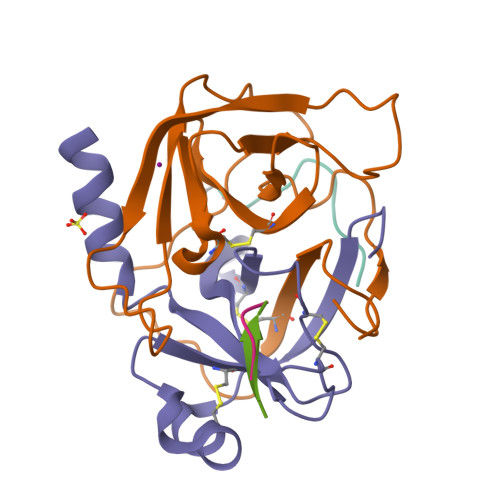
 PDB 5R4C | dpdb_5cuug8krCrystal Structure of gamma-Chymotrypsin at pH 9, room temperatureUpdated Oct 21, 2026
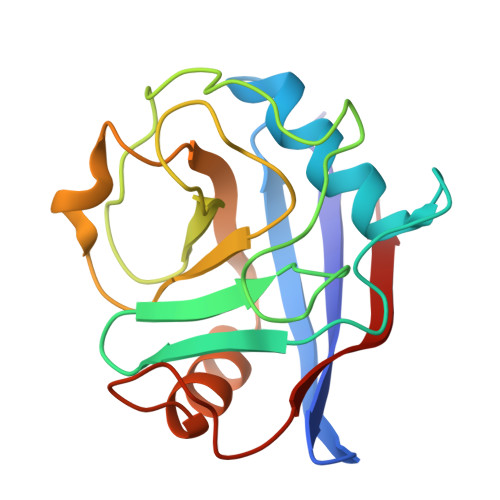 PDB 3QYU | dpdb_cd75ivw0Crystal structure of human cyclophilin D at 1.54 A resolution at room temperatureUpdated Oct 6, 2026
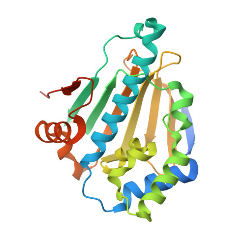 PDB 7S8Z | dpdb_9mq8l30nRoom-temperature apo Human Hsp90a-NTDUpdated Oct 6, 2026
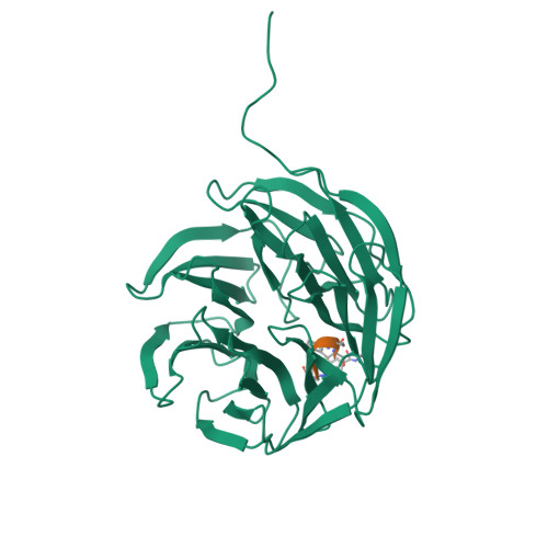 PDB 7AXX | dpdb_1d6atnhvStructure of WDR5:CS-VIP8 crystal after illumination at 405 nm and room temperatureUpdated Oct 6, 2026
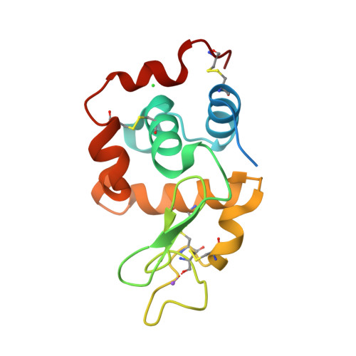 PDB 7DTF | dpdb_5rljgu8rRoom temperature structure of lysozyme by serial millisecond crystallographyUpdated Oct 5, 2026
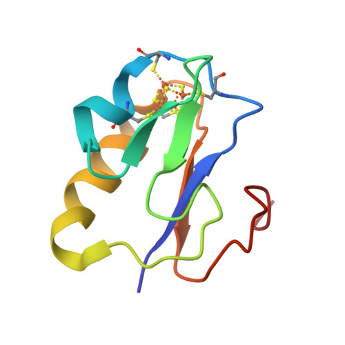 PDB 7YL8 | dpdb_ycfmq4lvNeutron structure of Bacillus thermoproteolyticus Ferredoxin at room temperatureUpdated Oct 5, 2026
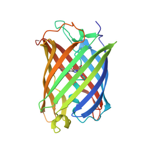 PDB 7TSR | dpdb_r44e5c8iRoom temperature rsEospa Cis-state structure at pH 8.4Updated Oct 5, 2026
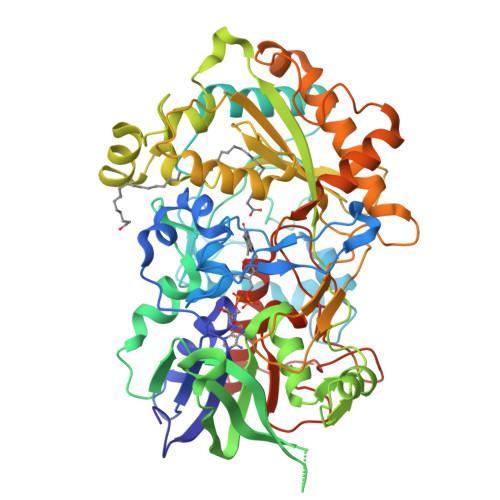 PDB 6YRX | dpdb_pbncowp7Low-dose crystal structure of FAP at room temperatureUpdated Oct 5, 2026
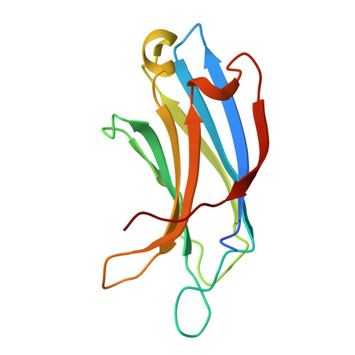 PDB 8FAF | dpdb_64gf8nikUnedited Octopus bimaculoides Synaptotagmin 1 C2A at room temperatureUpdated Oct 5, 2026
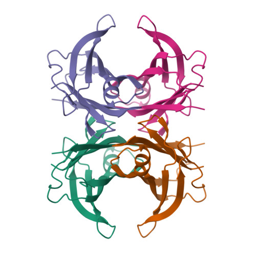 PDB 4PVL | dpdb_evsda652X-ray structure of human transthyretin (TTR) at room temperature to 1.9A resolutionUpdated Oct 5, 2026
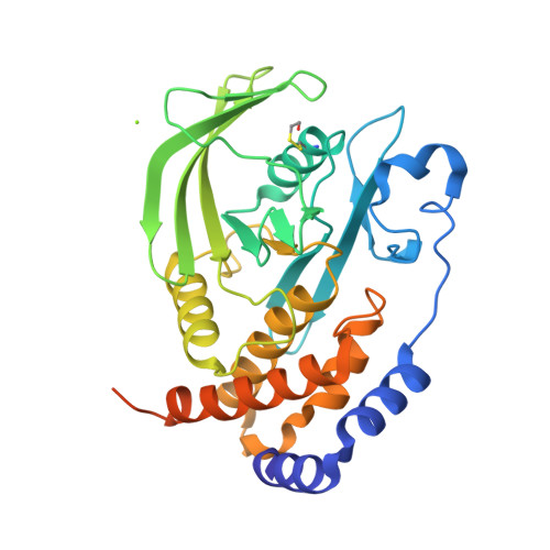 PDB 9CYR | dpdb_wf8t3z5tCrystal structure of D245G mutant human PTP1B (PTPN1) at room temperature (298 K)Updated Oct 5, 2026
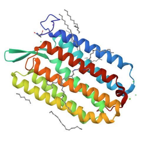 PDB 6S63 | dpdb_rmis8pylDark-adapted structure of Archaerhodopsin-3 obtained from LCP crystals using a thin-film sandwich at room temperatureUpdated Oct 5, 2026
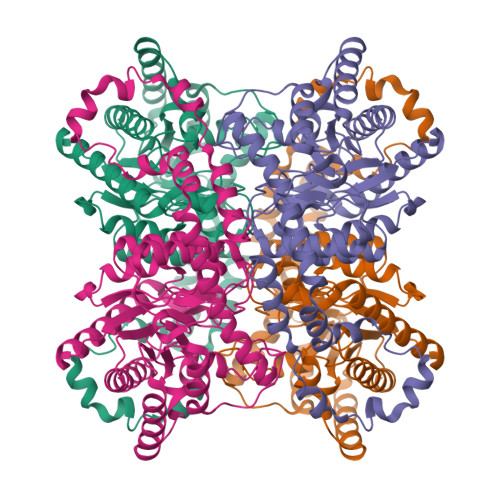 PDB 5I7G | dpdb_ovq7s011Metal free Glucose Isomerase collected at room temperature using the HC1b humidity controllerUpdated Oct 5, 2026
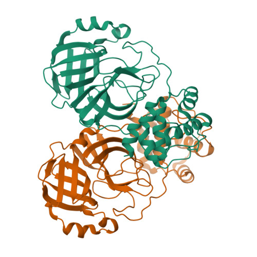 PDB 8E4J | dpdb_0m82jb1yRoom-temperature X-ray structure of SARS-CoV-2 main protease H41A miniprecursor mutantUpdated Oct 5, 2026
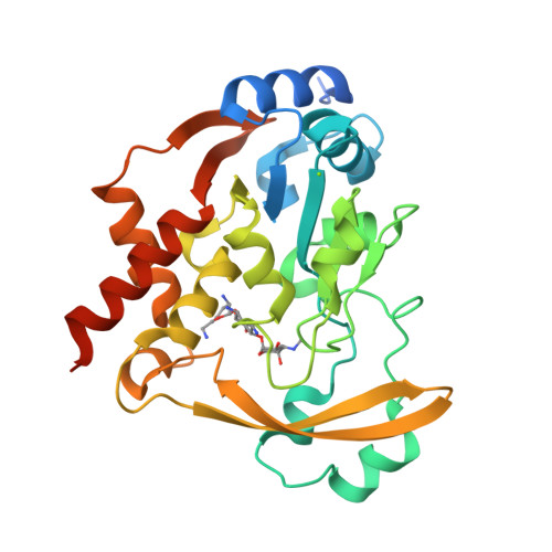 PDB 6BBZ | dpdb_k8ej72dgRoom temperature neutron/X-ray structure of sisomicin bound AAC-VIaUpdated Oct 5, 2026
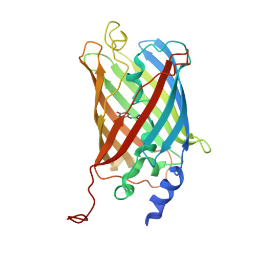 PDB 7O7V | dpdb_0za7xbetCrystal structure of rsEGFP2 mutant V151A in the fluorescent on-state determined by serial femtosecond crystallography at room temperatureUpdated Oct 5, 2026
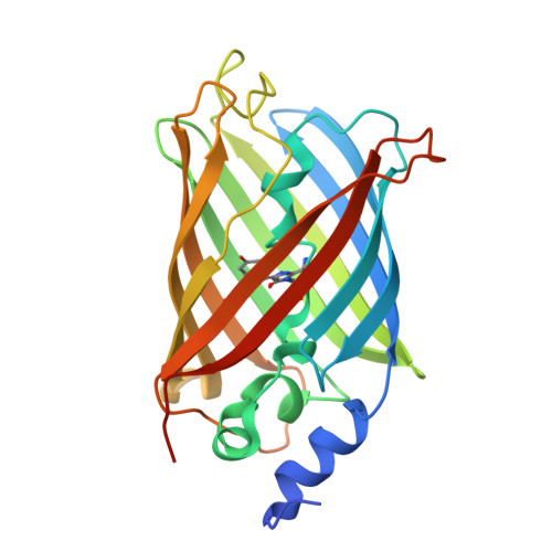 PDB 7O7X | dpdb_0yoth8csCrystal structure of rsEGFP2 mutant V151A in the non-fluorescent off-state determined by serial femtosecond crystallography at room temperatureUpdated Oct 5, 2026
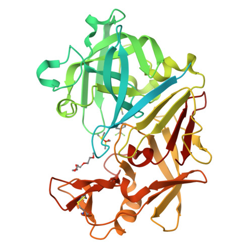 PDB 7QM9 | dpdb_ahj4gmjuEndothiapepsin in complex with compound TL00150 at room-temperature (temperature ramping down structure 1)Updated Oct 5, 2026
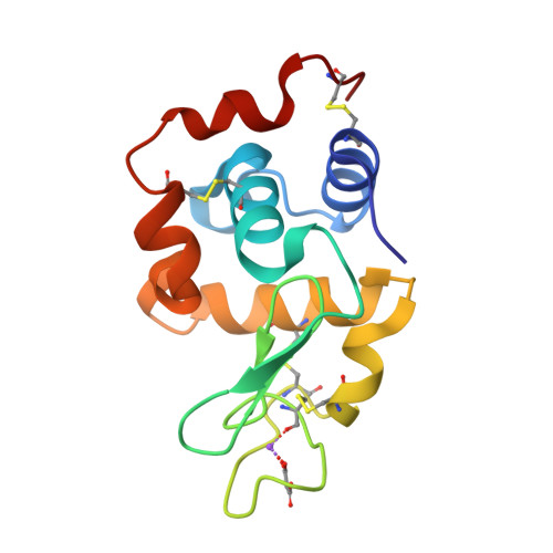 PDB 7CDP | dpdb_2imi7dgyLysozyme room-temperature structure determined by SS-ROX combined with HAG method, 42 kGy (3000 images)Updated Oct 5, 2026
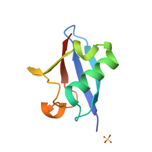 PDB 5TOF | dpdb_s749cupiRoom temperature structure of ubiquitin variant u7ub25Updated Oct 4, 2026
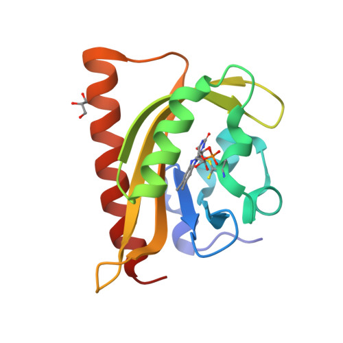 PDB 2V1B | dpdb_updbuo7kN- and C-terminal helices of oat LOV2 (404-546) are involved in light-induced signal transduction (room temperature (293K) light structure of LOV2 (404-546))Updated Oct 4, 2026
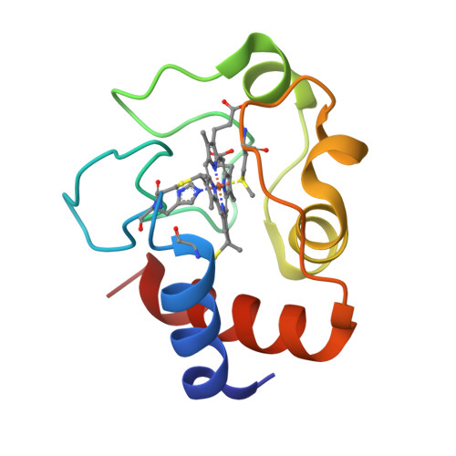 PDB 6K9I | dpdb_58scrnhaHigh-resolution three-dimensional structure of horse heart cytochrome C at room temperatureUpdated Oct 4, 2026
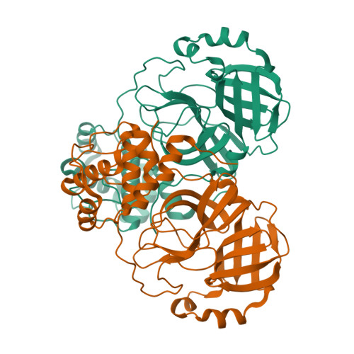 PDB 6XHU | dpdb_nxczy8x9Room temperature X-ray crystallography reveals oxidation and reactivity of cysteine residues in SARS-CoV-2 3CL Mpro: Insights for enzyme mechanism and drug designUpdated Oct 4, 2026
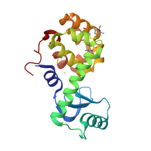 PDB 5G27 | dpdb_ayay8topStructure of Spin-labelled T4 lysozyme mutant L118C-R1 at Room TemperatureUpdated Oct 4, 2026
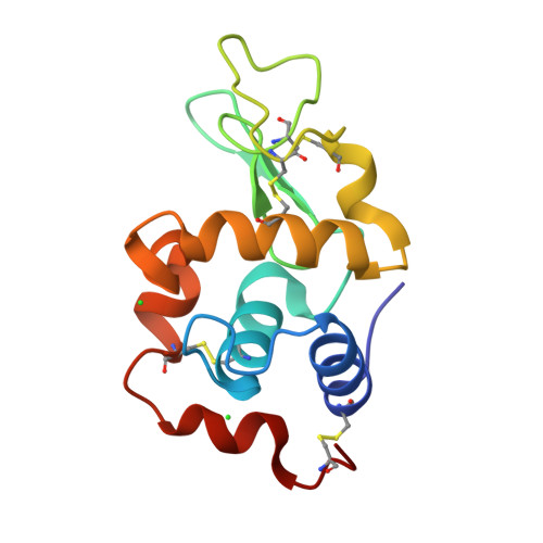 PDB 3ZEK | dpdb_p3ysplexHen egg-white lysozyme structure determined at room temperature by in- situ diffraction in ChipXUpdated Oct 4, 2026
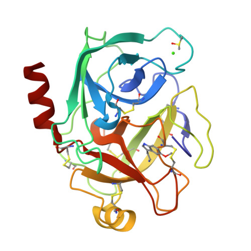 PDB 7WB8 | dpdb_6h09aisdCrystal structure of Bovine Pancreatic Trypsin in complex with 5-Methoxytryptamine at Room TemperatureUpdated Oct 4, 2026
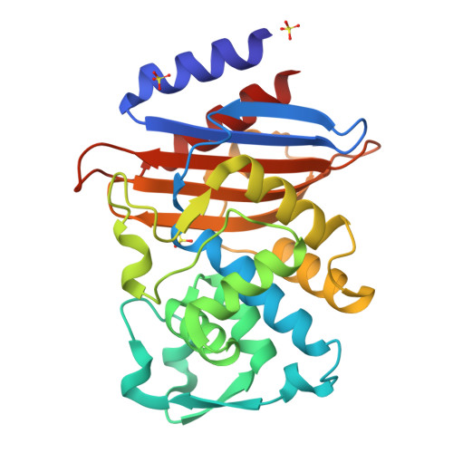 PDB 8AF8 | dpdb_wr5qgdfkRoom temperature SSX crystal structure of CTX-M-14 (5K dataset)Updated Oct 4, 2026
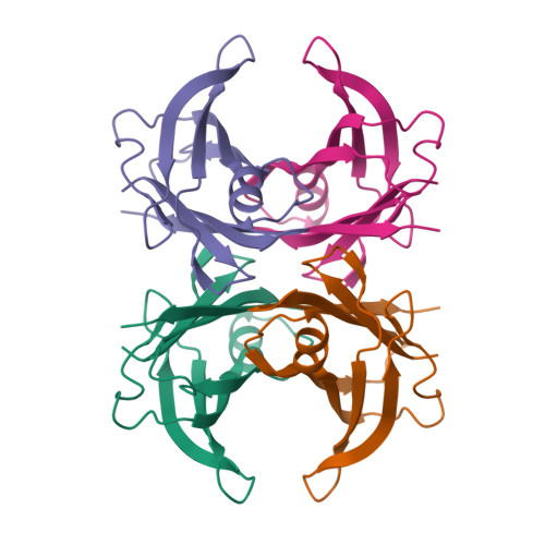 PDB 5NFW | dpdb_7f7dh2plNeutron structure of human transthyretin (TTR) S52P mutant at room temperature to 1.8A resolution (quasi-Laue)Updated Oct 4, 2026
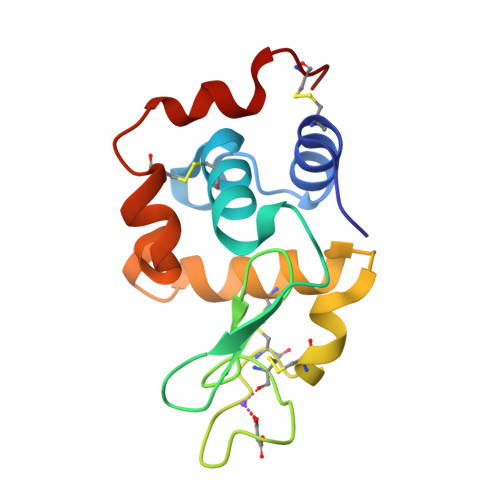 PDB 7CDK | dpdb_d055wlvaLysozyme room-temperature structure determined by SS-ROX combined with HAG method, 42 kGy (4500 images from 1st half of data set)Updated Oct 4, 2026
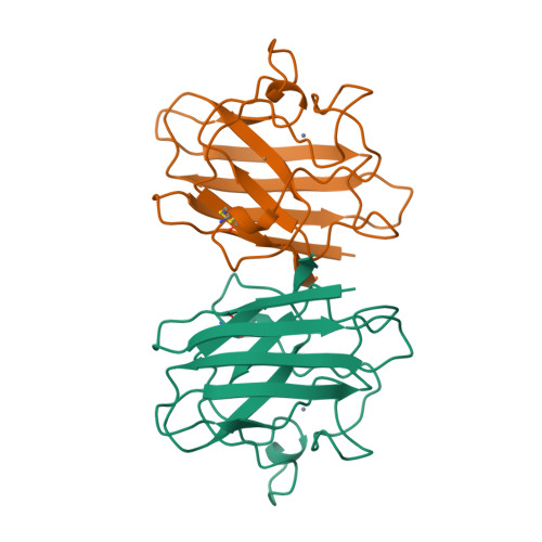 PDB 1B4L | dpdb_kmedtdgb15 ATMOSPHERE OXYGEN YEAST CU/ZN SUPEROXIDE DISMUTASE ROOM TEMPERATURE (298K) STRUCTUREUpdated Oct 4, 2026
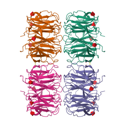 PDB 7B7F | dpdb_y29ltjuzRoom temperature X-ray structure of H/D-exchanged PLL lectin in complex with L-fucoseUpdated Oct 4, 2026
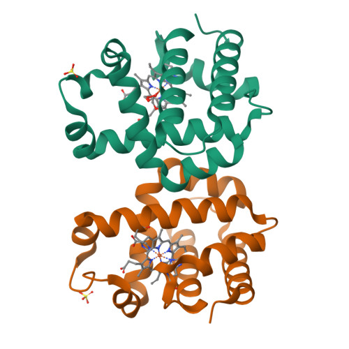 PDB 7KFM | dpdb_253nxvl4Room temperature oxyferrous Dehaloperoxidase BUpdated Oct 4, 2026
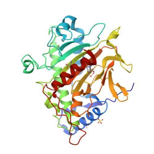 PDB 6ZAI | dpdb_vqgumc41Room temperature XFEL Isopenicillin N synthase structure in complex with Fe, O2 and ACV after exposure to dioxygen for 1600ms.Updated Oct 4, 2026
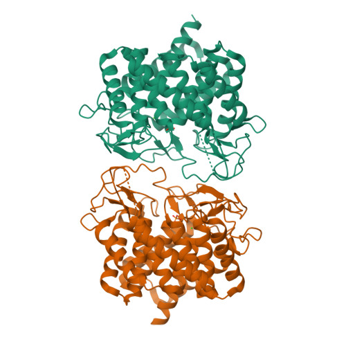 PDB 8BRY | dpdb_14uwzxosRoom-temperature structure of Pedobacter heparinus N-acetylglucosamine 2-epimerase at atmospheric pressureUpdated Oct 4, 2026
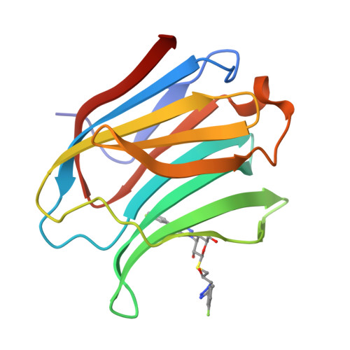 PDB 6RHL | dpdb_xrq0phwfRoom temperature data of Galectin-3C in complex with a pair of enantiomeric ligands: R enantiomerUpdated Oct 4, 2026
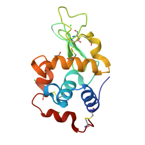 PDB 5LAN | dpdb_uhtp2n8mRoom temperature X-ray diffraction of tetragonal HEWL with 1M of uridine. Third data set (0.93 MGy)Updated Oct 4, 2026
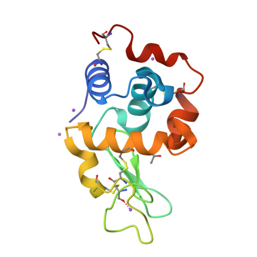 PDB 6QQ3 | dpdb_lb8xr23aThe room temperature structure of lysozyme via the acoustic levitation of a dropletUpdated Oct 4, 2026
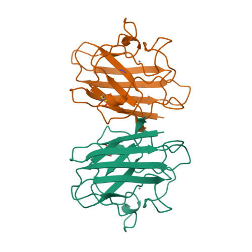 PDB 1YAZ | dpdb_q8mh0k03AZIDE-BOUND YEAST CU(II)/ZN SUPEROXIDE DISMUTASE ROOM TEMPERATURE (298K) STRUCTUREUpdated Oct 4, 2026
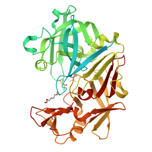 PDB 7QMC | dpdb_wufpq782Endothiapepsin in complex with compound TL00150 at room-temperature (temperature ramping down structure 4)Updated Oct 4, 2026
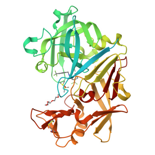 PDB 7QMO | dpdb_ei91jglxEndothiapepsin in complex with compound TL00150 at room-temperature (temperature ramping down structure 16)Updated Oct 4, 2026
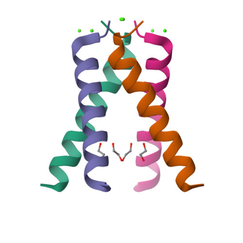 PDB 4QKL | dpdb_g15zo4jpInfluenza A M2 wild type TM domain at high pH in the lipidic cubic phase under room temperature diffraction conditionsUpdated Oct 3, 2026
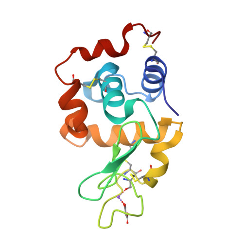 PDB 7CDO | dpdb_dvjq18zrLysozyme room-temperature structure determined by SS-ROX combined with HAG method, 21 kGy (3000 images)Updated Oct 3, 2026
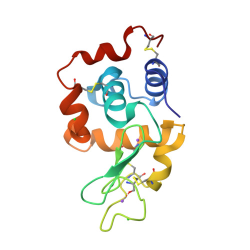 PDB 5FEK | dpdb_3cd1ey16Hen egg lysozyme at room temperature solved from 3600 diffraction images acquired by ultrasonic acoustic levitation method and processed by CrystFELUpdated Oct 3, 2026
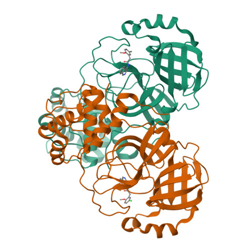 PDB 7S3K | dpdb_n7bildi1Room temperature X-ray structure of SARS-CoV-2 main protease in complex with compound Z1530718726Updated Oct 3, 2026
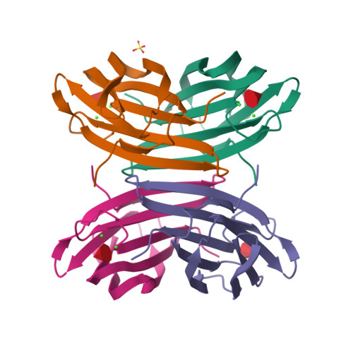 PDB 7PRG | dpdb_utu0v4ijJoint X-ray/neutron room temperature structure of perdeuterated LecB lectin in complex with perdeuterated fucoseUpdated Oct 3, 2026
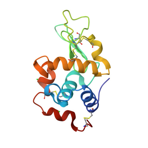 PDB 6QQF | dpdb_3nk2ivleRoom temperature structure of Hen Egg White Lysozyme recorded after an accumulated dose of 100 kGyUpdated Oct 3, 2026
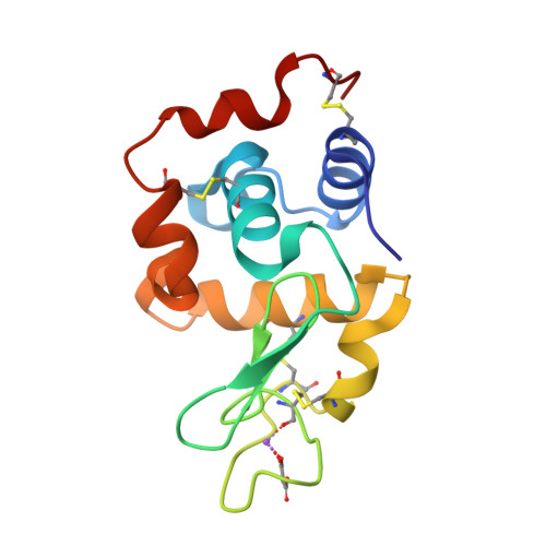 PDB 7CDM | dpdb_cqv4avbkLysozyme room-temperature structure determined by SS-ROX combined with HAG method, 42 kGy (4500 images from 2nd half of data set)Updated Oct 3, 2026
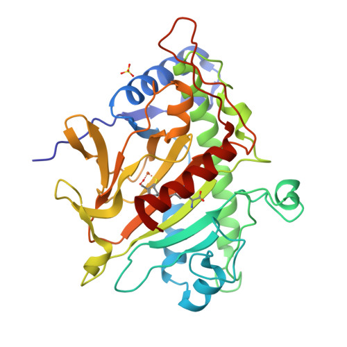 PDB 6ZAL | dpdb_9sn68qetRoom temperature XFEL Isopenicillin N synthase structure in complex with Fe and ACV after exposure to dioxygen for 500ms without glycerol.Updated Oct 3, 2026
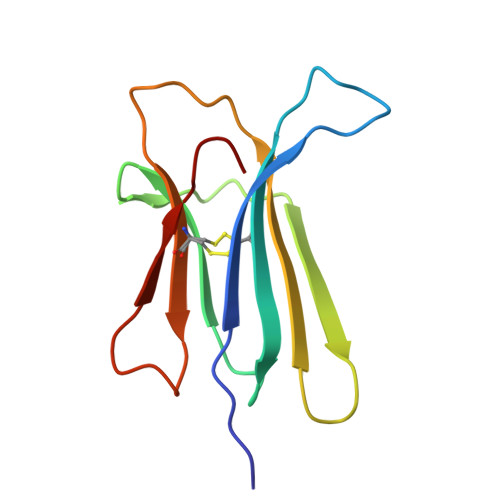 PDB 5CSB | dpdb_2bzvkr4jThe crystal structure of beta2-microglobulin D76N mutant at room temperatureUpdated Oct 3, 2026
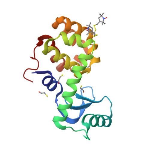 PDB 2OU8 | dpdb_4ur537waStructure of Spin-labeled T4 Lysozyme Mutant T115R1 at Room TemperatureUpdated Oct 3, 2026