
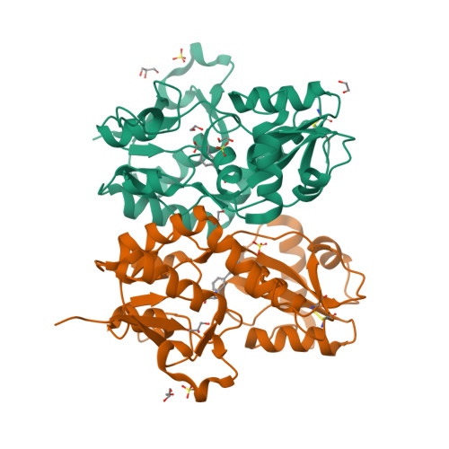
Rerefined model
PDB 5CBS|dpdb_3wq1i3a4
Rerefined model
Crystal structure of the GluA2 ligand-binding domain (S1S2J) in complex with the antagonist (R)-2-amino-3-(3'-hydroxybiphenyl-3-yl)propanoic acid at 1.8A resolution
- Program
- PHENIX 2.0_5824
- Model type
- Multiconformer
- Purpose
- Refinement
- Atoms
- 10,667
- Residues
- 1,042
- Protein chains
- 4
- Alternate-location fraction
- 1.54%
- Ligands
- CL, E42, EDO, GOL, SO4
Evaluations
R-work
0.168
R-free
0.216
Clashscore
4.5
Macromolecules
The same in every model of this entry.
Data
L0 Raw
L1 Processed
L2 Models
L3 Evaluations