
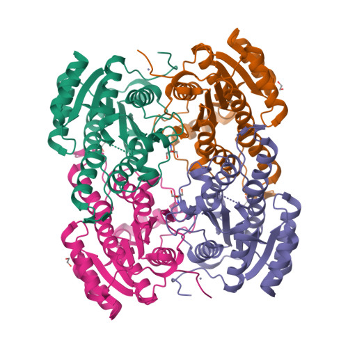
Deposited model
PDB 4O5O|dpdb_hsamxy0y
Deposited model
X-ray Crystal Structure of a 3-hydroxyacyl-CoA dehydrogenase from Brucella suis
- Program
- REFMAC 5.8.0049
- Model type
- Deposited
- Purpose
- Reference
- Authors
- Seattle Structural Genomics Center for Infectious Disease (SSGCID)
- Atoms
- 4,334
- Residues
- 518
- Protein chains
- 2
- Unmodeled fraction
- 1.52%
- Ligand
- ZN
Evaluations
R-work
0.136
R-free
0.169
Clashscore
2.4
Macromolecules
The same in every model of this entry.
| Entity 1 | |||||
|---|---|---|---|---|---|
| Molecule | Chains | Length | Organism | UniProt | Details |
| 3-hydroxyacyl-CoA dehydrogenase | 263 |
|
| — | |
Data
L0 Raw
L1 Processed
L2 Models
L3 Evaluations