
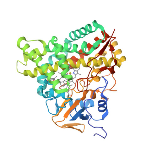
Deposited model
PDB 5X7E|dpdb_ozh1vw7k
Deposited model
Crystal structure of vitamin D hydroxylase cytochrome P450 105A1 (R84A mutant) in complex with 1,25-dihydroxyvitamin D2
CYP105A1 in complex with active form of vitamin D2
- Program
- REFMAC 5.7.0029
- Model type
- Deposited
- Purpose
- Reference
- Authors
- Hayashi, K., Yasuda, K., Shiro, Y., Sugimoto, H., Sakaki, T.
- Atoms
- 3,469
- Residues
- 404
- Protein chains
- 1
- Unmodeled fraction
- 1.94%
- Ligand
- HEM
Evaluations
R-work
0.186
R-free
0.232
Clashscore
3.3
Macromolecules
The same in every model of this entry.
Data
L0 Raw
L1 Processed
L2 Models
L3 Evaluations