
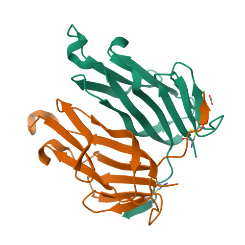
qFit model
PDB 3FC0|dpdb_rlxulxvp
qFit model
1.8 A crystal structure of murine GITR ligand dimer expressed in Drosophila melanogaster S2 cells
- Program
- Phenix 2.0_5824
- Model type
- Multiconformer
- Purpose
- Model Building
- Atoms
- 6,063
- Residues
- 242
- Protein chains
- 2
- Alternate-location fraction
- 98.35%
- Ligand
- ACT
Evaluations
R-work
0.167
R-free
0.217
Clashscore
—
Macromolecules
The same in every model of this entry.
Data
L0 Raw
L1 Processed
L2 Models
L3 Evaluations