
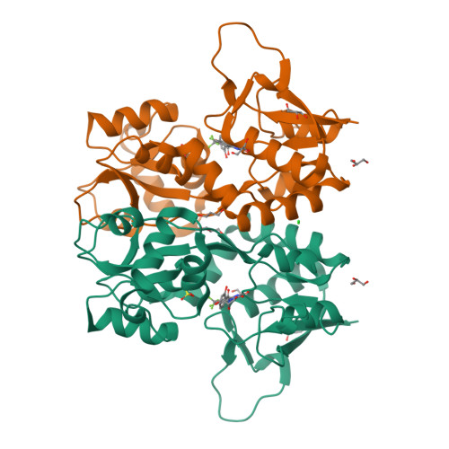
Rerefined model
PDB 6FZ4|dpdb_t5g0zd9t
Rerefined model
Structure of GluK1 ligand-binding domain in complex with N-(7-fluoro-2,3-dioxo-6-(trifluoromethyl)-3,4-dihydroquinoxalin-1(2H)-yl)-2-hydroxybenzamide at 1.85 A resolution
- Program
- PHENIX 2.0_5824
- Model type
- Multiconformer
- Purpose
- Refinement
- Atoms
- 2,424
- Residues
- 250
- Protein chains
- 1
- Alternate-location fraction
- 11.2%
- Ligands
- CL, EC8, GOL, SO4
Evaluations
R-work
0.197
R-free
0.243
Clashscore
5.3
Macromolecules
The same in every model of this entry.
Data
L0 Raw
L1 Processed
L2 Models
L3 Evaluations