
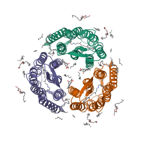
Deposited model
PDB 6G7J|dpdb_u4oi91ds
Deposited model
Retinal isomerization in bacteriorhodopsin revealed by a femtosecond X-ray laser: 457-646 fs state structure
- Program
- PHENIX (1.11.1_2575
- Model type
- Deposited
- Purpose
- Reference
- Authors
- Nogly, P., Weinert, T., James, D., Cabajo, S., Ozerov, D., Furrer, A., Gashi, D., Borin, V., Skopintsev, P., Jaeger, K., Nass, K., Bath, P., Bosman, R., Koglin, J., Seaberg, M., Lane, T., Kekilli, D., Bruenle, S., Tanaka, T., Wu, W., Milne, C., White, T., Barty, A., Weierstall, U., Panneels, V., Nango, E., Iwata, S., Hunter, M., Schapiro, I., Schertler, G., Neutze, R., Standfuss, J.
- Atoms
- 2,018
- Residues
- 230
- Protein chains
- 1
- Unmodeled fraction
- 12.21%
- Ligand
- RET
Evaluations
R-work
0.205
R-free
0.264
Clashscore
2.7
Macromolecules
The same in every model of this entry.
Data
L0 Raw
L1 Processed
L2 Models
L3 Evaluations