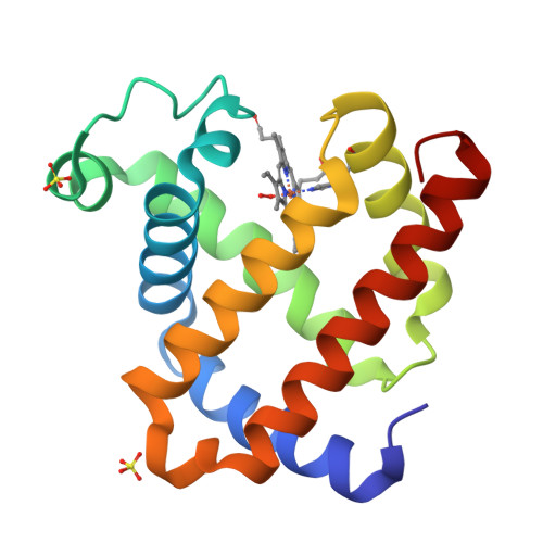
qFit model
PDB 2Z6S|dpdb_vbkr0fyu
qFit model
Crystal structure of the oxy myoglobin free from X-ray-induced photoreduction
- Program
- Phenix 2.0_5824
- Model type
- Multiconformer
- Purpose
- Model Building
- Atoms
- 2,197
- Residues
- 151
- Protein chains
- 1
- Alternate-location fraction
- 52.98%
- Ligands
- HEM, OXY, SO4
Evaluations
R-work
0.152
R-free
0.176
Clashscore
—
Macromolecules
The same in every model of this entry.
Data
L0 Raw
L1 Processed
L2 Models
L3 Evaluations