
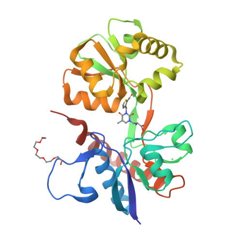
qFit model
PDB 2F34|dpdb_yz15qcrb
qFit model
Crystal Structure of the GluR5 Ligand Binding Core Dimer with UBP310 At 1.74 Angstroms Resolution
- Program
- Phenix 2.0_5824
- Model type
- Multiconformer
- Purpose
- Model Building
- Atoms
- 7,816
- Residues
- 502
- Protein chains
- 2
- Alternate-location fraction
- 55.18%
- Ligands
- 1PE, CL, UBA
Evaluations
R-work
0.189
R-free
0.226
Clashscore
—
Macromolecules
The same in every model of this entry.
Data
L0 Raw
L1 Processed
L2 Models
L3 Evaluations