
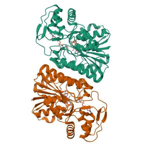
qFit model
PDB 7YSM|dpdb_zgsaipau
qFit model
Crystal Structure of UDP-glucose 4-epimerase (Rv3634c) co-crystallized with UDP-N-acetylglucosamine from Mycobacterium tuberculosis
- Program
- Phenix 2.0_5824
- Model type
- Multiconformer
- Purpose
- Model Building
- Atoms
- 9,041
- Residues
- 622
- Protein chains
- 2
- Alternate-location fraction
- 54.18%
- Ligands
- ACT, NAD, UD2
Evaluations
R-work
0.149
R-free
0.200
Clashscore
—
Macromolecules
The same in every model of this entry.
Data
L0 Raw
L1 Processed
L2 Models
L3 Evaluations